Monday, January 29, 2018

Congenital Heart Disease part 7


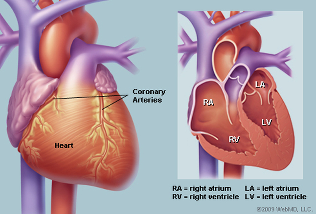
PULMONARY ATRESIA WITH VENTRICULAR SEPTAL DEFECT

Pulmonary atresia with ventricle septal defect, sometimes also called tetralogi of Fallot with pulmonary atresia, shares the intracardiac anatomy of tetralogy but lacks a direct communication between the right ventricle and pulmonary artery.  Patients with pulmonary artery and ventricle septal defect have a good size right ventricle, and from this perspective are suitable biventricular repair, major problems with pulmonary artery are common and determine both clinical presentation and management (the complexity of the pulmonary vascular bed may make repair unattractive or impossible).

Early clinical presentation varies between cyanosis (for patients with reduced or threathened pulmonary blood flow), failure to thrive, and/or exertional dyspnoea, to heart failure (for patients with execessive pulmonary blood flow through large major aortic pulmonary collaterals, who may develop segmental pulmonary arterial hypertension with time).

When pulmonary blood flow is is duct dependent, profound cyanosis and cardiovascular collapse ensue as the duct closes. Patients with confluent, good size pulmonary arteries and a pulmonary trunk (usually with valvar atresia) are suitable for a Fallot-like repair using  a trans annular patch. Patients with good sized pulmonary arteries but without  a pulmonary trunk should undergo repair with a right ventricle-pulmonary arteries conduit. Patients with confluent but hypoplastic pulmonary arteries often require an arterial shunt or reconstruction of the right ventricle outflow tract (without  ventricle septal defect closure), which may enhance pulmonary arterial growth, and then be reviewed at a later stage for repair using a valved conduit. Patients with non-confluent pulmonary arteries with adequate, but not excessive, pulmonary blood flow in infancy can survive into adulthood without surgery. There are proponents of a staged unifocalization approach for this latter challenging group of infants, ultimately aiming for a conduit repair.

Late clinical presentation for repaired patients is similar to those with tetralogy, whereas unrepaired patients present with exertional dyspnoea, fatigue and progressive chronic cyanosis, the latter leading to multi organ involvement and, with time, to a number of complications:
  • Hemoptysis may be due to rupture of usually small collateral vessels and/or pulmonary artery thrombosis
  • Chronic heart failure is usually multi factorial and may be due to chronic cyanosis, early excessive pulmonary blood flow, increased pulmonary vascular resistence, right ventricle dysfunction, aortic regurgitation
  • Progressive dilatation of the ascending aorta with increasing aortic regurgitation and aortic disecction (very rare complication) may occur
  • Endocarditis can be particularly compromising in patients with limited cardiovascular reserved and those with significant cyanosis
  • Increasing cyanosis may be due to decreased pulmonary blood flow from collateral stenosis, pulmonary artery stenosis, increased pulmonary vascular resistance or increasing ventricular end diastolic pressure
  • Arrhytmia and sudden cardiac death are not uncommon

Congenital Heart Disease part 6


EBSTEIN'S ANOMALY

Ebstein's anomaly is a relative rare disease and characterized by abnormally formed and apically displaced  leaflets of the tricuspid valve. Tricuspid valve opening is dislocated away from from the tricuspid valve annulus towards the apex or the right ventricular outflow tract. The anterior leaflet usually originates appropriately at the annular level but enlarged and sail-like, while the septal and posterior leaflet are displaced towards the right ventricular apex and often tethered to the endocardium.

The apical displacement of the tricuspid valve means that the right heart consists of an right atrium, an atrialized portion of the right ventricle, and the remaining functional right ventricle. The tricuspid valve is often regurgitant.

The most frequently associated anomalies include a shunt at the atrial level (secundum atrial septal defect or patent foramen ovale) and accessory pathways (Wolf-Parkinson-White syndrome). Ventricle septal defect, pulmonary stenosis, pulmonary atresia, tetralogy of Fallot, coarctation of the aorta, or mitral valve abnormalities can also occur .

Ebstein's anomaly occurs more commonly if the mother has received lithium or benzodiazepins during pregnancy.

The morphological and haemodynamic spectrum is wide. Haemodynamic changes depend on the severity of the tricuspid valve dysfunction, the degree of atrialization of the right ventricle, contractility of the remaining functional and the systemic ventricle, type and severity of concomitant anomalies and arrythmias.

The pathophysiology is characterized by systolic regurgitation of blood from the functional right ventricle, across the tricuspid valve, into the atrialized ventricle or right atrium, which tend to dilate. An interatrial connection permits an left-right shunt, or especially during exercise, an right-left shunt. Eibstein anomaly may result in chronically low systemic cardiac output.

The clinical presentation ranges from trivial symptoms to the presentation of profound cyanotic heart
defect. Patients with mild forms can be asymptomatic over decades until they are diagnosed. Key symptoms are arrhytmias, dyspnoea, fatigue, poor exercise tolerance, chest pain and peripheral and/or central cyanosis.


TETRALOGY OF FALLOT

Tetralogy of fallot is the most common form of cyanotic congenital heart diesease after 1 year of age, with an incidence approaching 10% of all forms of congenital heart defect. The defect is due to antero-cephalad deviation of the outlet septum resulting in the following four features:  a non-restrictive ventricle septal defect, overriding aorta (but<50%), right ventricular outflow  tract obstruction which may be infundibular, valvular, or (usually) a combination of both, with or without supravalvular or branch pulmonary artery stenosis, and consequent right ventricle hypertrophy.

Associated lesions include atrial septal defect, additional muscular ventricle septal defect, right aortic arch, anomalous (can be dual) left anterior descending coronary artery which may necessitate a conduit type of repair, and complete atrioventricular septal defect (rare, usually in association with down syndrome)

Early clinical presentation is dominated by a heart murmur in infancy and progressive cyanosis (from right to left  shunting at the ventricular level secondary to right ventricular outflow tract obstruction). Unoperated tetralogy of fallot carries a poor prognosis (>95% of patients used to die before 40 years of age). Early management may include paliative procedures to increase pulmonary blood flow.

Congenital Heart Disease part 5


RIGHT VENTRICULAR OUTFLOW TRACT OBSTRUCTION

Right ventricular outflow tract obstruction can occur at the subinfundibular, infundibular, valavular or supravalvular levels.

Subinfundibular stenosis commonly associated with a ventricle septal defect. It is caused by narrowing between prominent and hypertrophied muscle bands or ridges that separate the hypertrophied, high-pressure inlet and apical portions from a low-pressure, non-hypertrophied and non-obstructive infundibular portion of the right ventricle.

Infundibular stenosis usually occurs in combination with other lesions, particularly ventricle septal defec, tetralogy of fallot,and secondary to valvular pulmonary stenosis (reactive myocardial hypertrophy). At the infundibular level, and to some extent the subinfundibular level, the obstruction tends to be dynamic, meaning that the orifice narrows during systole.

Valvular pulmonary stenosis is usually an isolated lesions. Mainly due to intrinsic wall abnormalities and independent of haemodynamics, dilatation of the pulmonary trunk and the left pulmonary artery may occur, the right pulmonary artery generally being less affected. Most often, there is a typical dome-shaped pulmonary valved wit a narrow central opening but a preserved mobile valve base. A dysplastic pulmonary valve, with poorly mobile cusps and myxomatous thickening, is less common and frequenly part of Noonan syndrome. An hourglass deformity of the pulmonary valve, with bottle-shaped sinuses and stenosis at the comissural ridge of the valve, has also been described. In adults, a stenotic pulmonary valve may calcify late in life.

Supravalvular pulmonary stenosis or pulmonary arterial stenosis is caused by narrowing of the main pulmonary trunk, pulmonary arterial bifurcation, or pulmonary branches. It seldom occurs in isolation, and may occur in tetralogy of Fallot, Williams-Beuren syndrome, Noonan syndrome, Keutel syndrome, congenital rubella syndrome or Allagile syndrome. The stenosis may be located in the main branches or more peripherally. It may be discrete or diffuse (hypoplastic) or there may be frank occlusion, and may occur as single or multiple stenosis. Stenosis may be secondary to previous placement of a pulmonary artery band or at previous shunt site. A diameter stenosis >50%  is usually considered to be significant, and would be expected to have pressure gradient and a result in hypertension in the proximal pulmonary arteries.

Clinical presentation and natural hystory:
  • Subinfundubular/infundibular
Adults patient with unoperated may be asymptomatic or they may present with angina, dyspnoea, dizziness or syncope. The degree of obstruction is progressive over time.
  • Valvular
Patients with mild to moderate valvular pulmonary stenosis are usually asymptomatic. Mild valvular pulmonary stenosis in unoperated adults is usually not progressive. Moderate pulmonary stenosis can progress at the valvular level (calcification) or at the subvalvular, due to reactive myocardial hypertrophy. Patients with severe stenosis may present with dysnoea and reduced exercise capacity, and have a worse prognosis.
  • Supravalvular
Patients may be asymptomatic or have symptoms of dyspnoea and reduced exercise capacity. They are usually recognized  in the context of certain syndromes or referred for suspicion of pulmonary hypertension. Peripheral pulmonary artery stenosis may progress in severity.

Catheter intervention is recommended for patients with valvular pulmonary stenosis with valves which are not dysplastic (balloon valvotomy) and with peripheral pulmonary stenosis.
Surgery is recommended for patients with subinfundibular or infundibular pulmonary stenosis and hypoplastic pulmonary annulus, with dysplastic pulmonary valves.

Congenital Heart Disease part 4


COARCTATION OF THE AORTA

Coarctation of the aorta is considered as part of a generalized  arteriopathy, and not only as a circumscript  narrowing of the aorta. Its occurs as a discrete stenosis or as along, hypoplastic aortic segment. Typically coarctation of the aorta is located in the area where the ductus arteriosus inserts, and only in rare cases occurs ectopically (ascending, descending or abdominal aorta).

Associated lesions include bicuspid aortic valve, subvalvular, valvular or supravalvular aortic stenosis, mitral valve stenosis (parachute mitral valve, a complex known as Shone syndrome), or complex congenital heart defects. Coarctation aorta can be associated with Turner, Williams-Beuren, or congenital rubella syndromes, neurofibromatosis, Takayasu aortitis or trauma.

Coarctation aorta imposes significant afterload on the left ventricle, resulting in increase wall stress, compensatory left ventricular hypertrophy, left venticle dysfuctions, and the development of arterial collaterals.

Cystic medial necrosis with early elastic fibre fragmentation and fibrosis was found in the ascendings and decendings aorta, resulting in an increased stiffness of the aorta and carotid arteries.
Sign and symptoms depends on the severity of the coarctation of the aorta. Patients with serious coartation of the aorta exhibit signs and symptoms early in life, while particularly mild cases may not become evident until adulhood.

Key symptoms may include headache, nosebleed, dizziness, tinnitus, shortness of breath, abdominal angina, claudication, leg cramps, exertional leg fatigue and cold feet.

The natural course may be complicated by left heart failure, intracranial haemorhagge (from berry aneurysm), infective endocarditis, aortic rupture/dissection, premature coronary and cerebral artery disease, and associated heart disease.

In native coarctation of the aorta with appropriate anatomy, stenting has become the treatment  of first choice in adults in many centres. For adults with recurring or residual coarctation of the aorta, angioplasty with or without  stent implantation has been shown to be effective in experienced hands, and preferably stenting has also become first choice if anatomy is appropriate.

MARFAN SYNDROME

Marfan syndrome is an autosomal dominant disorder of connective tissue, in which cardiovascular, skin and skeletal, ocular, pulmonary and dura mater abnormalities may be present to a highly variable degree.

Prognosis is mainly determined by progressive dilatation of the aorta, leading to aortic dissection or rupture, which are the major causes of death. The rate of dilatation is heterogeneous and unpredictable. The risk of type A dissecton clearly increases within increasing  aortic root diameter, but dissection may occasionally occur even in patients with only mild aortic dilatation. Other parts of the aorta may also be dilated . Patients with dilated aorta are usually asymptomatic. The presence of significant aortic, tricuspid, or mitral regurgitation may lead to symptoms of ventricular volume overload, but left ventricle disease may also occur independently.

Early identification and establishment of the diagnosis is critical, since prophylactic surgery can prevent aortic dissection and rupture.


Congenital Heart Disease part 3


LEFT VENTRICULAR OUTFLOW TRACT OBSTRUCTION

Left ventricular outflow tract obstruction can occur at the valvular, subvalvular, or supravalvular levels, isolated or at multiple levels.

VALVULAR AORTIC STENOSIS

The most common cause for congenotal valvular aortic stenosis is bicuspid aortic valve. Mutations in the NOTCH 1 gene have been related to bicuspid aortic valve. Abnormalities of the aortic wall which are associated with bicuspid aortic valve can lead to progressive dilatation, aortic aneurysm, rupture or dissection.

Patient frequently remain asymptomatic for many years. Progression of stenosis varies  and depends on initial severity, degree of calcification, age and atherosclerotic risk factors,  In bicuspid aortic valve, progression is faster in those patients with greater closure line eccentricity and an anteroposterior-oriented line of closure.
prognosis is good and sudden death is rare in asymptomatic patients with good exercise tolerance, even when stenosis is severe. Once symptoms (angina pectoris, dyspnoea or syncope) occur the prognosis deteriorates rapidly.

Symptomatic patients require urgent surgery. Medical treatment for heart failure is reserved only for non-operable patients. Neither statin treatment nor any other medical treatment has so far been shown to retard progression of aortic stenosis.

SUPRAVALVULAR AORTIC STENOSIS

Supravalvular aortic stenosis can occur either as a localized fibrous diaphragm just distal to the coronary artery ostia or, most commonly, as an external hourglass deformity with a corresponding luminal narrowing of the aorta, or as diffuse stenosis of the ascending aorta. It fequently occurs as part of the Williams-Beuren syndrome, and may be associated with hypoplasia of the entire aorta, involvement of coronary ostia or stenosis of major branches of the aorta or pulmonary arteries.

Patients present with symptoms of either outflow obstruction or myocardial ischaemia. Sudden death occurs rarely, but it is more common in supra aortic stenosis with Williams-Beuren syndrome, with diffuse peripheral pulmonary artery stenosis, or with coronary artery disease. Surgery is the primary treatment.

SUBAORTIC STENOSIS

Subaortic stenosis can occur as an isolated lesion, but is frequently associated with a ventricle septal defect, an atrioventricular septal defect, or Shone complex, or may develop after correction of these lesions. It caused by a fibrous ridges in the left ventricular outflow tract proximal to the aortic valve or as fibromuscular narrowing and has to be differentiated from hypertophic cardiomyopathy.

The clinical course is highly variable. Aortic regurgitation is frequent but rarely haemodynamically significant or progressive.

Surgical treatment involves a circumferential resection of the fibrous ring and parts of the muscular base along the left septal surface.

Congenital Heart Disease part 2


ATRIOVENTRICULAR SEPTAL DEFECT

An atriventricular septal defect (Atrioventricular canal or endocardial cushion defect) is characterized by the presence of a common antrioventricular annulus, guarded by five leaflets. In the partial form, the anterior and posterior bridging leaflets are fused centrally, creating separate left and right sided orifices. In the complete atrioventricular septal defect the central fusion is not present and there is only one orifice. The partial atrioventricular septal defect (Primum atrio septal defect, partial atrioventricular canal) has a defect only at the atrial level. A complete atrioventricular defect (Complete atrioventricular canal) has a septal defect in the crux of the heart, extending into both the interatrial and interventricular septum. The atrioventricular node is positioned posterior and inferior to the coronary sinus. The bundle of His and the left bundle branch are displaced posteriorly. This accounts for an abnormal activation sequence of the ventricles.

Clinical presentation will mainly depend on the presence and size of the atrial septal defect and ventricle septal defect and competence of the left sided atrioventricular valve. Symptoms are not specific for an atrioventricular septal defect  and are caused bay intracardiac shunting (left-right, right-left, or bidirectional), pulmonary hypertension, atrioventricular  valve regurgitation, ventricular dysfunction or left ventricular outflow tract obstruction. Exercise intolerance, dyspnoea, arrhytmia and cyanosis may be present. Subaortic stenosis may be present or develop in time.

The history of unoperated complete atrioventricular septal defect is that of Eisenmenger syndrome unless the ventricle septal defect  is only small.

Unrepaired partial atrioventricular septal defect is not uncommon in adults. The presenting clinical symptoms are that of an left-right shunt at the atrial level and/or that of left-sided atrioventricular valve regurgitation. Patient may be still asymptomatic , but symptoms tend to increase with age. Most adults are symptomatic by 40 years of age.

PATENT DUCTUS ARTERIOSUS

Patent ductus arteriosus is the persistent communication between the proximal left pulmonary artery and decending aorta just distal to the left subclavian artery. It can be associated with a variety of congenital heart disease lesions. However, in the adult it is usually an isolated finding.

Patent ductus arteriosus originally result in left-right shunt and left ventricle volume overload. In moderate or large patent ductus arteriosus, pulmonary pressure is elevated. In patients who reach adulthood with a moderate patent ductus arteriosus, either left ventricle volume overload or ppulmonary arterial hypertention may be predominant (genetic predisposition). Adult patients with large patent ductus arteriosus have in general Eisenmenger physiology.

Presentations of adult patients with patent ductus arteriosus include:
  • Small duct with no left ventricle volume overload (normal left ventricle) and normal pulmonary artery pressure (generally asymptomatic)
  • Moderate patent ductus arteriosus with predominant left ventricle volume overload, large left ventricle with normal or reduced function (may present with left heart failure)
  • Moderate patend ductus arteriosus with predominant pulmonary arterial hypertention, pressure overloaded right ventricle (may present with right heart failure)
  • Large patent ductus arteriosus, Eisenmenger physiology with differential hypoxaemia and differential cyanosis (lower extrimities cyanotic, sometimes left arm too)

Congenital Heart Disease part 1


ATRIAL SEPTAL DEFECT:

Atrial septal defect may not uncommonly remain undiagnosed until adulthood.
Atrial septal defect types include:
  • Secundum atrial septal defect (located in the region of the fossa ovalis and its surrounding)
  • Primum atrial septal defect (partial atriaventricular septal defect, partial atrioventricular canal, located near the crux, atrioventricular valves are typically malformed resulting in various degrees of regurgitation
  • Superior sinus venosus (located near the superior vena cava entry, associated with partial or  complete connection of right pulmonary veins to superior vena cava, right atrium)
  • Inferior sinus venosus defect (located near the inferior vena cava entry)
  • Unroofed coronary sinus (separation from the left atrium can be partially or completely missing)
Associated lesions include anomalous pulmonary venous connection, persistent left superior vena cava,  pulmonary valve stenosis  and mitral valve prolapse.
The shunt volume depends on right ventricle/left ventricle compliance, defect size, and left atrial/right atrial pressure. A simple atrial septal defect results in left to right shunt because of the higher compliance of the right ventricle compared with the left ventricle (relevant shunt in general with defect size > 10mm), and causes right ventricle volume overload  and pulmonary overcirculation. Reduction in left ventricle compliance or any condition with elevation of left atrial pressure (hypertention, ischaemic heart disease, cardiomyopathy, aortic and mitral valve disease) increase left to right shunt. Reduced right ventricle compliance (pulmonic stenosis, pulmonary arterial hypertention, other right ventricle disease) or tricuspid valve disease may decrease left to right shunt or eventually cause shunt reversal resulting in cyanosis.
Patients frequently remain asymptomatic until adulthood. However, the majority develop symptoms beyond the fourth decade including reduced functional capacity, exertional shortness of breath and palpitations (supraventricular tachyarrhythmias) and less frequently pulmonary infections and right heart failure.

VENTRICULAR SEPTAL DEFECT

As an isolated finding,  ventricular septal defect is the most common congenital heart malformation at birth, if bicuspid aortic valve is not counted. It is mostly diagnosed and -when indicated- treated before adulthood. Spontaneous closure is frequent. Several locations of the defect within the interventricular septum are possible and can be divided into four group :
  • Perimembranous/paramembtranous/conoventricular (located in themembranous septum with possible extension into inlet, trabecular or outlet septum; adjacent to tricuspid and aortic valve. aneurysms of the membranous septum are frequent and may result in partial or or complete closure)
  • Muscular/trabecular (completely surrounded by muscle, various locations, frequently multiple, spontaneous closure particularly frequent)
  • Outlet supracristal/subarterial/subpulmonary (located beneath the semilunar valves in the conal or outlet septum; often associated with progressive aortic regurgitation due to prolapse of the aortic cusp)
  • Inlet/Atrioventricular canal (inlet of the ventricular septum immediately inferior to the atrioventricular valve apparatus, typically occuring in Down Syndrome)
Often there ios single defect, but multiple defects do occur. Ventricle septal defect is also a comnmon component of complex anomalies such as tetralogy of fallot, transposition of the great arteries etc.  Spontaneous closure of a ventricle septal defect can occur.

The direction and magnitude of the shunt are determined by pulmonary vascular resistance, the size of the defect, lef ventricle/ right ventricle systolic and diastolic function, and the presence of right ventricular outflow tract obstruction.

Therapeutic consideration for grown-up congenital heart disease


With exceptions, medical management is largely supportive for heart failure, arrythmias, pulmonary and systemic arterial hypertension, prevention of thrombo-embolic events, or endocarditis. Significant structural abnormalities usually require interventional treatment.

Heart failure:

Heart failure is a frequent problem in the grown-up congenital heart disease population.  However, as the pathophysiology of cardiorespiratory dysfunction is often very different from the failing normal circulation,  particularly in settings such as transposition of the great arteries with arterial switch repair

Arrhythmias and sudden cardiac death:

Arrhytmias are the main reason for the hospitalization of grown-up congenital heart disease patient and they are an increasingly frequent cause of morbidity and mortality. Furthermore, the onset of arrythmyas may be a signal of haemodynamic decompensation, and the risk associated with arrhytmias may be amplified in the presence of the often abnormal underlying circulation.
Sudden cardiac death is of particular concern in grown-up congenital heart disease. The  defects with the greatest known risk of late sudden cardiac death are tetralogy of fallot, transposition of the great arteries, aortic stenosis and univentricular heart. Unexplained syncope is an alarming event.

Surgical treatment:

Many grown-up congenital heart disease patients will have undergone intervention in childhood, but surgery during adulthood may be required in various situation:
  • Patients with prior repair and residual or new haemodynamic complication
  • Patient with conditions not diagnosed or not considered severe enough to require surgery in childhood
  • Patients with prior palliation

Catheter intervention:

There has been a marked increase in the number and range of interventional catheterization procedures in grown-up congenital heart disease, which in some patients obviates the need for surgery. In others, treatment of congenital cardiac malformations is best achieved by a collaborative approach involving interventional catheterization and surgery.  Newer techniques include stenting of systemic or pulmonary vessels and percutaneous valve implantation.

Infective endocarditis:

The endocarditis ris in grown-up congenital heart disease patients is substantially higher than in the general population, with marked variation between lesions. The approach to antibiotic endocarditis prophylaxis has changed for several reason. In short, transient bacteraemia occurs not only after dental procedures but frequently in the context of daily routine activities such as tooth brushing, flossing or chewing. The recommendation is limited to dental procedures requiring manipulation of the ginggival or periapical region of the teeth or perforation of the oral mucosa.

Grown-up congenital heart disease


A thorough  clinical evaluation is of a critical importance in the diagnostic work up of  grown up congenital heart disease. The aim of analysing history is to assess present and past symptoms as well as to look for intercurrent events and any changes in medication. The patient should be questioned on his/her lifestyle to detect progressive changes in daily activity in order to limit  the subjectivity of symptom analysis. Clinical examination plays a major role and includes, during follow-up, careful evaluation with regard to any changes in auscultation findings or blood pressure or development of signs of heart failure. An electrocardiogram and pulse oxymetry are routinely carried out alongside clinical examination. Chest X-ray  is no longer performed routinely at each visit, but rather when indicated. It remains, nevertheless, helpful for long-term follow-up, providing information on changes in heart size and configuration as well as pulmonary vascularization.

Strategies for investigation of anatomy and physiology of congenital heart disease are changing rapidly, with a shift from invasive studies to non-invasive protocols involving not only echocardiography but, more recently, cardiovascular magnetic resonance and computed tomography.
Nuclear techniques may be required in special indications.

Evaluation of arrythmias, primarily in symptomatic patients.  Cardiopulmonary exercise testing has gained particular importance in the assessment and follow-up of grown-up congenital heart disease patients. It plays an important role in the timing of intervention or re-intervention.

Diagnostic work-up:
  • Echocardiography
Echocardiography remains the first line investigation and continuous to evolve, with improved functional assessment using four dimensional echocardiography, doppler tissue imaging and its derivatives, contrast echocardiography and perfusion imaging transoesophageal echocardiography.
Echocardiography provides, in most instances, information on the basic cardiac anatomy including orientation and position of the heart, venous return, connections of the atria and ventricle,  and origin of the great arteries. It allows evaluation of the morphology of cardiac chambers, ventricular function, detection and evaluation of shunt lesions, as well as the morphology and functions of the heart valves. Assesment of ventricular volume overload (increase in end-diastolic volume and stroke volume) and pressure overload (hypertrophy, increase in ventricular pressure) is of major importance. Doppler echocardiographic information also includes  haemodynamic data such as gradient across obstructions and right ventricle pressure/pulmonary artery pressure , but also flow calculations.  Although echocardiography can provide comprehensive information, it is highly user dependent.
  • Cardiac magnetic resonance imaging
Cardiac magnetic resonance imaging has become increasingly important in grown-up congenital heart disease patients and is an essential facility in the specialist unit. It enables excellent three dimensional anatomical reconstruction, which is not restricted by body size or acoustic windows and has rapidly improving spatial and temporal resolution. It is particularly useful for volumetric measurements, assessments of vessels, and detection of myocardial fibrosis.
  • Computed tomography
Computed tomography plays an increasing role in imaging of grown-up congenital heart disease patients,  providing excellent spatial resolution and rapid aquisition time. It is particularly good for imaging epicardial coronory arteries and collateral arteries, and for parenchimal lung disease. Ventricular size and function can be asssessed, with inferior temporal resolution compared with cardiac magnetic resonance imaging. The drawback of most current computed tomography systems is its high dose of ionizing radiation , making serial use unattractive.
  • Cardiopulmonary exercise testing
Cardiopulmonary exercise testing , including assessment  of objective exercise capacity (time, maximum oxygen uptake), ventilation effeciency, chronotropic and blood pressure response, as well as exercise-induced arrythmia,  gives a broader  evaluation of fuction and fitness, and has endpoints which correlate well with morbidity and mortality in grown-up congenital heart disease.
  • Cardiac catheterization
Cardiac catheterization is now reserved for resolution of specific anatomical and physiological questions, or for intervention. Continuing indications include assessment of pulmonary vascular resistance, left ventricle and right ventricle diastolic function, pressure gradients,  and shunt quantification when non-invasive evaluation leaves uncertainty, coronary angiography, and the evaluation of extracardiac vesseels such as aortic pulmonary collateral arteries.

Therapeutic cardiac catheterization with congenital heart defect


Valvuloplasty or balloon valvotomy:

This procedure is done to open a narrowed heart valve. Any of the heart's four valves can be narrowed. However, this procedure is most often used to open the valves connecting the heart to the lungs (pulmonary valve) or to the body (aortic valve). These narrowing occur bacause the valve leaflets don't open up completely. This makes it harder for the heart to pump blood to the lungs or to the body, The narrower the valve, the more pressure it takes to pump the blood through it.  To open the narrowed valve, a special catheter with a balloon attached to its end is used.
A picture of the valve is first taken and the size of the valve is measured carefully to select the correct-size balloon. If the ballon to small, the opening still not be big enough. If the ballon is to large, the valve may be damaged or the vessel may be torn. The balloon is inflated for only a few seconds, then it is deflated and removed.

Angioplasty:

This procedure widens a narrowed blood vessel. These narrowings are often associated with various congenital heart defects and can occur naturally or after surgery, Similar to a narrowed valved, a narrowed vessel restrict blood flow and causes the heart to work harder. Blood vessels that can be narrowed include:
  • The branch pulmonary artery
  • The aorta
  • Systemic veins
  • Pulmonary veins
Stent Implantation:

Sometimes, simply widening a narrowed blood vessel with balloon isn't effective. The narrowing in the vessel may be too long or it might strech out with the balloon but shrink again once the balloon is removed.

In this situation, a stent is used to provide structural support within the narrowed vessel to keep it wide open. Stent are metal mesh tubes. They are designed to strech open inside a narrowed blood vessel and hold the vessel wall open.

There are many types of stent, but the most common ones used in children are "balloon -expandable" stents. These are mounted onto a balloon and positioned at the site of narrowing through a long sheath. Then the balloon is inflated to expand the stent against the narrowed  vessel wall. The stent is opened to the appropriate size depending on the patient's size. The balloon is deflated and removed while the stent stays in the vessel to keep it from  renarrowing.

Balloon and blade septostomy:

In some special circumstances, it's necessary to create a larger hole between the walls of the heart's upper chambers (the right and left atrium). Special balloons and blade catheters are used to create  these openings to increase blood flow between the heart's upper chambers. This procedure can be performed in the cath lab or by the bedside in the intensive care unit under ultrasound guidance.

Valve perforation:

Some patients are born with a completely blocked pulmonary valve. This is called pulmonary atresia. When this occurs and blockage is due to a thin membrane of the valve, the blocked valved can be opened in the cardiac cath lab using radiofrequency perforation. This technique uses a special catheter that can generate heat to create a small opening in the blocked valve. Then the catheter can be placed across the valve and the opening enlarged using the same technique as described in the valvuloplastysection. When the blockage is due to a thickened abnormal valve, it may not be feasible to make an opening and surgery might be needed.

Occlusion procedure:
These procedure are used to plug up (or close) an unwanted opening or connection in the heart or in blood vessels. These heart defects can be closed using a variety of devices.

Therapeutic cardiac catheterizations for children with congenital heart disease


A therapeutic cardiac catheterization is a procedure performed to treat heart defect. A doctor will use a special techniques and a thin plastic spaghetti-like tube or catheter that goes to the heart from blood vessels in the legs or the neck. These techniques allow the repair to be done without surgically opening the chest and heart. The types of defects that can be repaired include closing a hole in the wall that seperates the heart's right and left sides, widening a narrowed vessel or stiff valve, and closing abnormal blood vessels using a variety of devices.

The doctor will decide whether your child willl receive sedation only or general anesthesia. Before entering the cardiac catheterization laboratory (cath lab), a medication may be given to help your child relax and fall asleep.

If sedation chosen, your child will also have an IV started. This allows medications to be given to keep your child asleep and pain-free during the procedure. Your child will keep breathing on his own, and no breathing tube will be needed . Sometimes a topical numbing cream may be applied to both an IV site and to groin area where the catheters will be placed.

If the doctor chooses general anesthesia as best for your children, you will most likely meet with an anesthesiologist prior to the catheterization. Either an inhaled gas via a mask or an intravenous medication may be used for general anesthesia. After your child asleep, a breathing tube tube is inserted into the airway to make breathing easier. Additional medications and fluids may be given throughout the procedure to keep your child comfortable. When procedure is over, the breathing tube will be removed when your child is breathing on his own.

Catheterization is a sterile procedure performed using small catheters placed into blood vessels, so the risk of infection is minimal. Antibiotics aren't usually needed before or after the procedure. Removing the catheter, however, may cause blood to ooze into the skin. This can discolor the skin, like a black eye, but it doesn't usually cause problems.

The catheter doesn't hurt the heart and isn't painful once inside the heart, but its movement can cause abnormal heart rhythms. Your doctor can usually treat these rhythms by removing the catheter or using medications. Rarely, if the catheter touches the heart electrical system it can interfere with the spread of electricity. This is known as heart block. Although this is usually temporary, placing a special catheter connected to an electrical battery (pacemaker) may be required until the heart's electrical system corrects itself.

Tuesday, January 23, 2018

Postnatal congenital heart disease diagnosis


Cyanosis:

While babies with cyanotic heart disease may present blue at birth, in many is less obvious. The oxygen saturation has to be below 80% before cyanoses becomes clinically obvious. In some cyanotic conditions, the resting saturations will be in the 80's and so will not be obvious. Ductal dependent lessions eventually present with  rapidly progressive  cyanosis as the duct closes but the mixing situations may take some time to present. If there is any doubt about a baby, put a saturation monitor on the foot. If it is persistently less than 90%, further investigations should be initiated. The commonest condition presenting with neonatal cyanosis is transposition of the great vessels but any cyanotic congenital heart disease lesions can present in the newborn period.

Heart failure / respiratory distress:

It is unusual for the common left to right shunts to present in the newborn period because pulmonary pressure take longer to fall in the presence of a large left to right shunts. Typically, large ventricle septal defect   don't produce symptoms or signs in the neonatal period but present in failure at 2-4 weeks of age after the pulmonary pressures have fallen. Occasionally babies with left to right shunts will present with respiratory sign (usually tachypnoea), particularly if there is shunting at more than one site such as ventricle septal defect and patent ductus arteriosus or patent foramen ovale.

Shock / cardiovascular collapse:

This is the classic presentation of the ductal dependent obstructive left ventricle conditions (hypoplastic left heart, critical aortic stenosis and coarctation). In these conditions, the only way blood can reach the systemic circulation is via the ductus. They are often asymptomatic while the duct is patent and then collapse any time during the first week when it closes. They present pale and shocked with respiratory distress and weak pulses.


INVESTIGATIONS

Echocardiography:

This is the definitive test and in situations where there is early access to these skills, most of the other investigations become superfluous. Early echocardiography to exclude congenital heart disease should be arranged in the following situations.

Chest Xray and electrocardiogram:

The traditional cardiac investigations of chest Xray and electrocardiogram are still useful in situations where there is limited access to echocardiography although both are limited use in excluding heart disease.

Hyperoxia test:

This is used for differentiating cardiac from non cardiac causes of cyanosis. Most babies with cyanotic heart conditions will not increase O2 pressure  significantly if placed in 100% oxygen.

Upper and lower limb blood pressure:

This maybe  useful in suspected coarctation although the accuracy of non-invasive blood pressure measures in babies is open to question. The difference between upper and lower limb blood pressures should be less than 15 mmHg.

Early detection of congenital heart disease


Congenital heart disease is one of the commonest human malformation.  It is  a clinical paradox that the most benign lesions such as small ventricle septal defect or mild pulmonary stenosis, are more likely to be detected on routine newborn examination. Of the major structural lesions, the cyanotic conditions will usually present with symptoms or signs and so also will be detected early. There remains however an important group with major structural lesions, particularly those with ductal dependent systemic circulations, who are well in the period shortly after birth and then collapse in a critical state once the ductus closes.

Risk factors of congenital heart disease:
  • Family history
  • Maternal diabetes
  • Other fetal abnormalities on  prenatal screening, including malformations of other systems, fetal arrythmia's and non-immune hydrops
  • Syndromes and other structural malformations diagnosed postnatally. Any baby with dysmorphic features or a structural malformation should be considered as at high risk for congenital heart disease. Congenital heart disease is a common in most chromosomal abnormalities and in many other non chromosomal syndromes, including fetal alcohol syndrome and congenital infection such as rubella.
  • Down syndrome

Antenatal diagnosis:

Babies will have had an ultrasound screening for malformations. This examinations usually includes a four chamber heart view. The use of the four chamber view also influences the type of lesions detected antenatally. Condition with major impact on chamber size  such as the hypoplastic left heart syndrome are more likely to be detected while conditions with little impact on chamber size such transposition or coarctation are less likely to be detected. Detection rates in high risk pregnancies are  much higher than for general screening and reflect the fact that referal is ussually made to a centre with expertise in fetal echocardiography.
Postnatal diagnosis:
  • An abnormal examination in an asymptomatic baby, usually a murmur
  • Cyanosis
  • Heart failure/respiratory distress
  • Shock/cardiovascular collapse
Abnormal clinical examination in an asymptomatic baby:
  • Assessement of colour
Many babies with cyanotic heart disease will be obviously blue from birth but some can be quite subtle, particularly with common mixing situations or ductal dependent pulmonary circulations before ductal constriction.  This is further confused by babies often having blue lips and extremities in the first day or two of birth. Look at the tongue can be helpful in babies with blue lips but, if in doubt, put an oxygen saturation monitor on the baby's foot.
  • Assessment of peripheral pulses including femorals
In post-ductal coarctation or preductal coarctation with a constricting  duct, weak femoral pulsess maybe the only clinical pointer. Unfortunately in the latter condition, while duct is still patent, the femorals maybe easily palpable. Feeling the femorals can be difficult in vigourous baby. One useful technique is to hold the leg in your hand and run your thumb up the medial border of the quadriceps muscle, your thumb will usually fall on the femoral pulse while your hand can limit the leg movement.
  • Assessment of the praecordial impulses
While there is no rigorous study of the accuracy of this sign, many major congenital heart disease lessions will produce an increased right and/or left venticular load well before a murmur appears. So an easily palpable or visible praecordial impulse in a quite baby should always be taken seriously as a possible sign of congenital heart disease.
  • Ausculation for normal heart sounds and murmurs

The differentiation of dextroversion from dextroposition of the heart


Dextroversion  of the heart is a subgroup of dextrocardias, It includes the location of the heart in the right hemithorax without inversion of the cardiac chambers. This congenital anomaly  is the result of a counter-clockwise rotation of a normally developed heart in the right hemithorax. This condition has been described as an isolated phenomenon, as well  as associated with different intracardiac and/or pulmonary anomalies. The heart,  however,  may be positioned in the right hemithorax due to intrinsic factors, such as major pathology of the right lung or pleura.  There is no rotation of the heart in such cases, and the heart is simply drawn into the right hemithorax. This condition is mostly acquired and named dextroposition.

The terms dextroversion and dextroposition are sometimes interchanged, and different conditions are denoted  by the same name, or vice versa-different terms are used for the same condition.
The diagnosis of dextroversion of the heart is based on the clinical, roentgenologic, electrocardiographic or catheterization findings. On physical examination, in addition to the right-sided heart, the maximum impulse is felt near the sternum, and is caused by the anteriorly placed left ventricle and by the parasternal, anterior, ascending aorta. Abnormal auscultatory findings are present only when dextroversion is complicated by intracardiac anomaly.

In dextroposition of the heart normal cardiac chamber arrangement is present with more or less normal silhouette. The heart may be somewhat distorted by an underlying lung pathology.The apex of the heart is discernible to the left of the sternum or behind it, since the pulmonary or pleural pathology may pull the heart in toto, but does not rotate its apex and left ventricle anteriorly.
In dextroversion of the heart the electrogram finding extreme counterclockwise rotation of the heart with normally placed atria. The electrocardiogram in dextroposition of the heart does not show this type of ventricular rotation and approaches normal.

Cardiac catheterization is helpful in proving the normal position of the caval veins, the marked rotation of the ventricles by the right turn of the catheter from the right atrium into the right ventricle, and by the right to the left direction of the main pulmonary artery. These feature is not found in dextroposition of the heart.

All the above mentioned clinical, radiologic, electrocardiographic and catheterization findings may be changed and complicated by additional congenital anomalies of the heart or lungs, which though not infrequent-are not necessarily present.

Screening for critical congenital heart defect


Newborn screening for critical congenital heart defect may help identify newborns with these conditions and allow for timely care and treatment. Babies with a critical congenital heart defect usually need surgery or other procedures in the first year of life. Some babies born with a critical congenital heart defect appear healthy at first and may be sent home with their families before their heart defect  is detected.  These babies  are at risk for having serious complications within the first few days or weeks of life and often require emergency care.

Typically, critical congenital heart disease lead to low levels of oxygen in a newborn and maybe identified using pulse oximetry screening at least 24 hours after birth. Once identified, babies with a critical congestive heart disease can be seen by heart doctors (cardiologists) and can receive specialized care and treatment that could prevent death or disability early in life.

About 1 in every 4 babies born with a heart defect  has a critical congenital heart defect. Babies with a critical congenital heart defect are at increased risk for death or disability if their condition is not diagnosed soon after birth.

Newborn screening for critical congenital heart defect involves  a simple, painless, bedside test called pulse oximetry in wich sensors are placed on the baby's skin. This test measures the amount of oxygen in a baby's blood. Low levels of oxygen in the blood can be a sign of a critical congenital heart defect.

Critical congenital heart defect :
  • Coartation of the aorta
  • d-Transposition of the great artery
  • Double-outlet right ventricle
  • Ebstein anomaly
  • Hypoplastic left heart syndrome
  • Interrupted aortic arch
  • Pulmonary atresia (with intact septum)
  • Single ventricle
  • Total anomalous pulmonary venous connection
  • tetralogy of fallot
  • Tricuspid atresia
  • Truncus arteriosus

Thursday, January 18, 2018

Fetal cardiac evaluation by 3D/4D ultrasonography


The congenital heart disease are the most common major malformation at birth.  In 2003, with the development of Spatio-Temporal Image Correlation (STIC), scientists started the use of third and fourth dimension ultrasonography (3D/4D) in fetal cardiac evaluation. The STIC is a software that enables acquiring volumetric fetal heart with  its vascular connection, whose images can be evaluated  either multiplanar or rendering modes, or even surface mode, in a static or moving ways (4D) by means of cineloop, which simulates a complete cardiac cycle.Thus, a detailed assesment of the anatomy and the functioning fetal heart is possible without the need to cause major discomfort to pregnant  women.

Standarization of volume storage is already a reality. so the investigator has knowledge of the actual position of the heart chambers with respect to the right and left fetal axis to evaluate the presence of possible cardiac isomerism. Therefore,  when the fetus is in cephalic presentation, it should be considered that the heart side corresponds to the fetal side, unlike the pelvic fetuses which stay in opposite sides.

The gray scale and color Doppler applications are also present in the STIC, used to improve the evaluation of the ventricular outflow tracts, aortic and ductal arches, besides assisting in the location of septal defect. The 3D technology has allowed the development of new techniques known as inversion mode (analysis technique of liquid  structure which reverses voxels of gray scale, so anechoic structures such as the heart chambers and lumen vessels, with inversion mode they become echogenic and B-flow imaging (technique that improves the weak signal reflected from the blood, and supresses the strong signals of the surrounding structures). The inversion mode allows the reconstructionof the cardiac chambers, aortic and ductal arches, and abnormalities of venous connection. The B-flow imaging shows high sensitivity and angle independence, then it is potentially advantageous over colour Doppler for the visualization of large vessels and venous return, allowing the identification of small vessels with low-velocity flows, such as pulmonary veins, enhancing the detection of anomalies in pulmonary venous return.

Another technique also addressed by STIC Tomographic Ultrasound Imaging's (TUI), which enables the achievement of all the axial planes of the heart from the abdoment to the apex of the chest, increasing the fetal heart tracking and analysis. STIC also allows the measurement of volume cardiac chambers, as well as the calculation of systolic volume, ejection fraction and cardiac output. Thus, to obtain relevant information about cardiac function due to the congenital heart disease. More recently, a new approach to STIC rendering mode analysis obtained measurements of mitral and tricuspid valves areas, and also the interventricular septum, determining reference values for these parameters, making it feasible to apply in suspected or pathological cases.

Ultrasonography


High frequency sound waves are used to obtain images of the unborn baby and the different structures of the baby. These sound waves pass through a device known as tranducer, which then pass through the amniotic fluid surrounding the baby and bounce off the baby harmlesly, creating "echoes". These "echoes" are converted into images by the computer and screened on the monitor so that the outline of the baby and the internal structures can be seen. Currently, the equipment used are known as real-time scanner, whereby continuous image of the moving fetus can be seen on the monitor screen. It has become a very useful diagnostic tool during pregnancy.


Ultrasonography for pregnancy    :
  • Confirm pregnancy
As early as five weeks of gestation, the gestational sac can be seen on an                ultrasound. At six weeks, the embryo can be measured and observed.  The scan also helps to confirm the number of babies.
  • Determine age and size of fetus
Measurements of different part of the body reflect the age  and size of the fetus. This is particularly important in early gestation. In patients with uncertain last menstrual periods,  such measurements must be made as early as possible in pregnancy to arrive at a correct dating for the patient (dating scan). During later part of pregnancy, these measurements help in assesing growth of the fetus (growth scan)
  • Diagnosis of fetal malformation
Ultrasound scanning allows comprehensive surveys of the fetal anatomy to detect the presencen of structural anomalies of the brain, heart, kidneys, lungs, limbs and other organs. Physical abnormalities in the fetus and fetal organs  can often  be detected via ultrasound within 22 weeks of pregnancies (fetal anomaly scan)
  • Placental localisation
An ultrasound scan is very useful in identifying the site of the placenta. This assists the physician in excluding a placenta praevia (placenta lying close to the neck of the womb) and other placental abnormalities.
  • Doppler blood flow studies
This is a special type of scan  which allows the physician to study in great details the blood flow to various fetal organs and the placenta.
  • Other diagnosis
Ultrasound scan can be used to confirm fetal presentation, evaluate fetal movements,  tone and breathing, determine the fetal gender and diagnose uterine and pelvic abnormalities during pregnancy such as fibroids and ovarian cyst.

Types of ultrasound scan
  1.   Transvaginal         :   Scan is done through  the vagina
  2.   Transabdominal   :   Scan is done through the mother's abdoment

Ultrasound


Recent advances in prenatal diagnosis and therapy has been made possible with the invention of newer imaging modalities including 3D and 4D ultrasound. Two dimensional ultrasound remains the method by which most fetal structural abnormalities are screened and diagnosed, however 3D and 4D are being used increasingly for the examination of the human fetus. Two dimensional scanning allows visualisation of static images while 3D and 4D imaging adds a further dimension to fetal study by allowing interaction with volume data sets to examine anatomic structures of interest in planes of section.

The examination of fetal face by 3D ultrasound has generated a great deal of intereest by both the medical fraternity and prospective parents. The "photographic-like" images are easily recognized by both the layperson and expert alike. Facial expressions such as mouth-opening, tongue protursion and yawning may be studied in detail using 4D ultrasound. This has lead several  investigators to hypothesise that the adjuctive use of 3D/4D ultrasound would improve the diagnostic accuracy of 2D ultrasound. The advantage of 3D ultrasound was an improvement  in the diagnostic accuracy to detect clefts of the palate and decrease in the number of false positive diagnoses.

The benefits of 3D ultrasound in fetal  central nervous system include:
  • The ability to determine the severity, location and extent of central nervous system abnormalities
  • The possibility of reconstructing and visualising the corpus callosum in the sagital plane from volume data sets
  • The ability to visualise  the  3 horns of the ventricular system in a single plane

The ability to visualise the level of defect in cases of spina bifida using 3D imaging is important in counselling regarding prognosis and treatment.
Conditions such as diapraghmatic hernia, skeletal dysplasias and preterm premature rupture of membranes are associated with risk of pulmonary hypoplasia. Prognosis is dependent on the residual size of the affected lung.
The fetal heart should be examined in motion but with standard 3D the spatio and temporal resolution of the image is limited. It is further complicated by motion artefacts.

Development of the human external ear


External ear development is a lengthy snd complex precess that extends from early embryogenic life until well into the postnatal period. Initial development of the auricle and external auditory canal during the fourth and fifth weeks of gestation is closely associated with anotomycal changes involving the pharingeal arch apparatus of the human embryo. The auricle and external canal are well formed by the time of birth but do not attaintheir full size and adult configuration until about 9 years of age. Sebaceous and modified apocrine glands, which are responsible for cerumen production, begin their development at about 5 months gestation in association with hair follicles in the outer portion of the external canal. Although they appear anatomically mature before birth, these glands do not reach full fuctional capacity until puberty.

As emphasized throughout this special issue, the external ear plays an essential role in auditory function and occupies an important place in the clinical practice in audiology and otology. Its major component, the auricle and external auditory canal, receive sound energy from the environtment and provide some degree of directional and frequency selectivity for the incoming sound and stimulus. They also serve to protect the tympanic membrane from mechanical injury and from abrupt changes in temperature and humidity. Various abnormalities affecting the external ear, particularly those involving congenital defects, are best understood from a developmental perspective.

External ear development is a process that begins in embryonic life, progresses through the fetal period to the time of birth, and continoues postnatally until the age of puberty, when the glands of the external canal become fully functional. (As ussually defined, the embryonic phase of human development extends from 2 weeks gestational age up to the seventh or eight week, while the fetal period is the interval from about 8 weeks gestation to term).

The embryonioc pharingeal arch apparatus provide the structural foundation for formation of the external ear.  The pharingeal arches are conspicious external feature of the human embryo and are significantly involved in various aspects of head and neck development.

By the end of the forth week of gestation, four well-defined pairs of pharingeal arches are externally visible in the neck region of the human embryo. The first two of these, the mandibular and hyoid arches, are important contributors to external ear development. During the fifth gestational week, nodular swellings of tissue known as the hillocks of his appear on the first and second pharingeal arches. Six such hillocks , three on either side of the first pharingeal cleft, can be distinguished. Most investigators believe that the auricle is formed by growth, differentiation and fusion of these six tissue condensation. During the initial stages of its development, the auricleis located in the general area of the neck, behind the lower jaw, but by the 20th week of gestation it has moved upward to attain its adult location and overal configuration. In a 4-5 years old child, the auricle is about 80 percent adult size. It reaches full adult size by approximately 9 years of age.

Brain Development in Children


Clothes baby

Early childhood is the most and rapid period of development in a human life. The years from conceptions through birth to eight years of age critical to the complete and healty cognitive, emotional and physical growth of children.

A Child's brain develops in response to both genes and the environtment. It is the interaction between the genes and the environtment that really shape the developing brain, a dance between biology and experience.While the gene provide the initial map for development, it is the experiences and relationships babies and children have every day that literally shape their brains. Families have an extremely important ongoing influence on children's development. The community and service environtments in which children and families interact also play a key role in supporting optimal development.

The rapid development of children's brains begin in the prenatal stage and continous after birth. Although cell formation is virtually complete before birth, a new born baby has about a 100 billion brain cells, brain maturation, and important neural pathway and connections are progressively developed after birth in early childhood. Therefore, early childhood is a period in development where environtment  actually has important impact on determaining how the brain and the central nervous system grows and develops, Environtment affects not only the number of the brain cells and the number of connections among them but also the way these connections are "wired". The process of eliminating excess neurons and synapses from the dense, immature brain, which continues well into adolescence, is most dramatic in the early yeras of life, and it is guided to large extent by the child's sensory experience of the outside world. Scientific evidence suggest that if the brain doesn't receive the appropriate stimulation during this criticalwindow, it is very difficult for the brain to rewire itself at a later time.

Genes provide the initial map for brain development, beginning with the basic connections in the brain from birth. Significance wiring occurs during the first years of a child's life and this effectively programs child development. At three, a child has around 1000 trillion brain connections or synapses, which in later development are selectively pruned. When adolescence is reached, brain synapses will number around 500 trillion, and this number remains relatively stable into adulthood, The prunning of brain synapses indicates the tremendous influence experience and environtment play in shaping a young brain. It is the experiences and relationships that infants and young children have that continuosly develop their brains and build the neural circuits that will be the foundation for later development. New research in an area called epigenetics, even suggest that a person's genes can potentially develop in response to some environtmental factors.
Inadequate nutrition before birth and in the first years of life can seriously interfere with brain development and lead to such a neurological and behavioral disorders as learning disabilities and mental retardation. There is considerable evidence showing that infants exposed to good nutritions, and adequate psychosocial stimulation had measurably better brain function at twelve years of age than those raised in less stimulating environtment,

Stress is a feature of the normal development of positive and adaptive coping. Everyday stress responses of a moderate and brief nature can result in mild increases of hormone levels (cortisol) and short-lived increases in heart rate. These kinds of tolerable stress responses help in the development of adaptive coping when buffered by stable and supportive relationships and are an important part of healthy development.

Excessive or long -lasting stress is known as toxic stress and can have negative impact on brain development. Example of toxic stress include: physical or sexual abuse, neglect or lack of affection, parental mental illness, family violence, poverty and lack of adequate housing. Ongoing stress factors that are not buffered bay caring and positive relationships disrupt brain architecture leading to a lower treshold of activation of the stress management system, which in turn can lead to life long problems in learning , behaviour, and both physical and mental health,

Although manageable levels of stress are normal and growth promoting, toxic stress in the early years can damage brain development. It is in situations where ongoing stress is likely, that intervening as early as possible is critical to achieving the best possible outcomes for the child, Caring and positive relationships are essential to ensure stress levels promote resilience for babies and children.

The architecture of the brain (the neural circuits) is built in a hierarchical "bottom-up" sequence. This means the foundation is paramount, as higher level circuits are built on lower level ones. Each newly acquired skill aides in the sequential development of the next, Attaining the more complex and higher order skills becomes  much more difficult when the foundation is shaky. As the foundations are built upon, brain circuits stabilise making them much harder to change and this highlights the importance of getting them right the first time, Positive early experiences result in optimal brain development, which in turn provides the foundation for the other skills and abilities children need  for succes at school and for life.

These are critical periods, or "prime times" for various aspects of brain development. The brain is programmed for events and experiences to happen at particular times for the best wiring and brain development. For example, language developments depends on adequate hearing  and if hearing loss is not diagnosed at an early age and the brain can not receive the sounds that lead to language development, the language part of the brain begin to "close up". The quality of child's earliest environtments and the availability of appropriate expereiences at the right stages of development are crucial to brain development and the foundation for learning in later life,

Brain Development Period


ANTE NATAL:
  • All five senses begin to function before birth
  • Prenatal sensory experiences actually help shape the brain and nervous system
  • Prenatal experiences prime the attachment behaviours of the infant

0-3 YEARS:
  • A rapid periode of brain development which can be fostered by relationships with caregivers and supported by optimal community environtments for families and children
  • Brain development is vulnerable to toxic stress (depending on length and number of stressors for the child)

BY SCHOOL AGE:
  • Children build on the solid foundation of the first five years
  • It is more difficult for children to take advantage of the learning environtment of schools if they have not had an optimal home environtment, there is restricted access to quality early childhood sevices and they have experienced a poor quality community environtment

ADOLESCENCE
  • Brain development prioritises the connections used most often, Resulting in"pruning" of brain networks or circuits
  • As children entered this period, more intensive rsources are required if children have missed the opportunities for optimal caregiving and environtments in the preceeding years

Rethinking The Brain


Old thinking :
  • How the brain develops depens on the genes that you were born with
  • The experiences that you have before age three have a limited impact on later development
  • A secure relationship with primary caregiver creates a favorable context for early development and learning
  • Brain development is linier, the brain's capacity to learn and change grows steadily as an infant progesses towards adulthood
  • A toddler's brain is much less active than the brain of college student

New thingking :
  • How the brain develops hinges on a complex interplay between the genes that you are born wth and the experiences you have
  • Early experiences have decisive impact on the architecture of the brain, and on the nature and extent of adult capacities
  • Early interactions don't just create a context, they directly affect the way that the brain is wired
  • Brain development is non linear, there are prime times for acquiring different kinds of knowledge and skills
  • By the time children reach age three, their brains are twice as active as those of adults. Activity levels drop during adolescence

The brain reaches half its mature weght by about six months and 90 percent of its final weight by age eight. This rapid development is reflected in children's capabilities and what they do. Althoughevery children unique, it is widely accepted that development follows a basic pace and pattern of development in all children.

What Children Do at This Age


Baby


Birth to 3 months:
  • Begin to smile, tract people and objects with their eyes
  • Prefer faces and the bright colours
  • Turn toward sound
  • Discover feet and hands
 
4 to 6 months:
  • Smile
  • Develop preferences generally to parents and older siblings
  • Repeat actions with interesting results
  • Listen intently
  • Respond when spoken to
  • Laugh and gurgle
  • Imitate sounds
  • Explore hands and feet
  • Put objects in mouth
  • Sit when propped
  • Roll over
  • Grasp objects without using thumb
 
7 to 12 months :
  • Remember simple events
  • Identify themselves and body parts, and familiar voices
  • Understand their own name and other common words
  • Say first meaningful words
  • Explore objects and hidden objects
  • Put objects in containers
  • Sit alone
  • Put themselves up to stand and walk
 
1 to 2 years :
  • Imitate adult actions
  • Speak and understand words and ideas
  • Experiment with objects
  • Walk steadily, climb stairs and run
  • Recognize ownership of objects
  • Develop friendships
  • Solve problems
  • Show pride in accomplishments
  • Begin pretend play
 
2 to 3,5 years :
  • Enjoy learning new skills
  • Learn language rapidly
  • Gain increase control of hands and fingers
  • Act more independently
 
3,5 to 5 years :
  • Develop a longer attention span
  • Talk a lot, ask many questions
  • Test physical skills and courage with caution
  • Reveal  feeling in dramatic play
  • Like to play with friends, don't like to lose, share and take turns sometimes
 
5 to 8 years :
  • Gain curiosity about people and how the world works
  • Show more interest in numbers, letters, reading and writing
  • Gain more convidence and use words to express feelings and cope
  • Play cooperatively
  • Develop interest in final products

Prenatal Central Nervous System Development


Central Nervous System development plays the central role in the primary argument. That argument is that the prenatal nervous system is particularly vulnerable to environmenttal perturbations because it is rapidly developing during that time period. Futher, many of the learning and behavioral problems that occur in childhood and adolescence have their origins in these prenatal perturbation on central nervous system development.

To help conceptualize fetal central nervous system development, metaphorically link the development of the central nervous system to the construction of a house. In the same way that a blueprint guides house construction, an individual's genome serves as ablueprint for the brain. Some of the DNA in the genome creates proteins that buid structures, while others are"timing genes" that manage the sequencing of the building process, Neurons and glial cells function as the foundational materials of bricks, wood and cement. Axons, dendrites and synaptic connections among neurons serve as the wiring for electricity and the telephone.

The construction of this elaborate communication structure we call the brain is complex, but there are general principles that guide that process. First, while genes provide the blueprint, central nervous system development is a complex process that results from the interplay of genetically governed biological processess and a number of experential,environtmental factors. Second, despite this complex genetic and environtmental interaction, the formation of brain regions occurs according to precisety sequenced schedule with more phylogenetically primitive region (e.g, lymbic system, forebrain) developing before more complex structure (e.g, cerebral cortex). Third, within these regions, brain development is most vulnerable to insult during periods of most rapid growth and development. Thus, the timing of an insult may be more important than the dose or nature of the insult in fluencing the pattern of malformation. The fourth guiding principle is that of all organs in the body, the brain is most vulnerable to teratogenic disruptionbecause of the extended amount of time it requires for development. Fifth, birth does not mark a particular milestone in the development of the brain. The brain continues to develop throughout the lifespan, although the most significant development occurs early in the development during the fetal period and the first years of life.