Saturday, September 29, 2018

The central venous catheter part 7


Other key things to remember:
  • Sometimes central venous catheters can become blocked and will need medication to unblock them
  • Make sure the dressing stays dry and in place unless it is being changed by a nurse
  • Take a special care to keep the dressing dry when washing
  • Generally, you should only let the specially trained nephrology staff do anything with your catheter. In an emergency, other healthcare staff may need to use the catheter to get blood samples or to give medications

The central venous catheter part 6


At each hemodialysis treatment:
  • Make sure the nurses can see your central venous catheter at all times during your hemodialysis treatment
  • Wear a mask whenever the caps are removed from the ends of the catheter openings

The central venous catheter part 5


Tell your nephrology team right away if any of the following occur:
  • Fever, chills, sweats or tenderness on your skin at the exit site
  • New arm or facial swelling
  • Bleeding that occurs from your catheter or surrounding area when you are not in the dialysis unit

The central venous catheter part 4


On an ongoing basis you should:
  • Never remove the caps on the end of the central venous catheter openings
  • Avoid pulling on your catheter
  • Always wear a mask whenever the dressing is being changed and when the caps are being removed or replaced

The central venous catheter part 3


After central venous catheter placement:
  • Keep the dressing in place and dry at all times
  • Do not take a shower or soak the area in a bathtub
  • Do not swim
  • Report any fevers, chills or pain on your skin at the exit site to your nephrology team

The central venous catheter part 2


Placement procedure

The central venous catheter is put in using ultrasound to help with correct placement. A local anesthetic (freezing) is used.

The catheter is held in right place by stitch  on the skin. If your central venous catheter is going to be used permanently (because an arteriovenous fistula or graft cannot be made), the stitch may be removed once the catheter is firmly in place.

The central venous catheter part 1


The central venous catheter is a flexible synthetic (man-made) tube that is usually placed in a large vein in your chest. Sometimes these catheters are placed in a vein in the neck, groin or back. Each central venous catheter has two openings called "ports" or "limbs". One port takes blood from your body to be cleaned  by the dialysis machine and clean blood returns to your body  through the other port. The spot  where the catheter goes into the skin is called the "exit site.

A central venous catheter can be used while you are waiting for surgery for your arteriovenous fistula or graft. It is also an option if an arteriovenous fistula or graft cannot be made.

Central venous catheters are put in place by a nephrologist, surgeon or a radiologist in the x-ray department or the operating room.

A central venous catheter can be used immediately once it is placed in your vein. There is an increased risk of infection with central venous catheters at both area where the catheter goes into the body, and within the blood stream. Also, there may be damage to the vein where the catheter is placed.

Utility of intravascular ultrasound in peripheral intervention part 3


Intravascular ultrasound during intravascular stent delivery

Intravascular stents are currently used for many applications, including the prevention and treatment of complications associated with percutaneous transluminal angioplasty. Although stent has increased the patency rates of several endovascular procedures, it is not without risks. Many of the complications are related to inappropriate selection of lesions for treatment with stents, or maldeployment of stents. Inadequate stent expansions can lead to early thrombosis or stent migration, and over-expansion of the stent can result in excessive intimal hyperplasia or vessel perforation. Intravascular ultrasound has been particularly useful for stent placement in both coronary and peripheral lesions.

Three-dimensional reconstruction is particularly useful to assess the completeness of the stent expansion. On the reconstructed images of the stented vessel, the stent structure can be seen as independent of the arterial walls. Thus, incomplete stent implantation is evident. The incorporation of an intravascular ultrasound transducer into the stent delivery system will permit simultaneous imaging during stent delivery, enhancing the precision of the procedure.

Utility of intravascular ultrasound in peripheral intervention part 2


Intravascular ultrasound has been shown to provide a useful perspective during percutaneous balloon angioplasty. Before intervention, intravascular ultrasound has been used to discriminate fibrous from calcified plaques and concentric  from eccentric lesions. Following procedures, intravascular ultrasound has accurately detected the presence of dissections and plaque fractures, and has shown that in such cases luminal enlargement after transluminal balloon angioplasty is produced by stretching of the arterial wall while the volume of the lesion remain constant.

In addition to specific plaque characteristics, several other variables, such as luminal thrombus, extensive dissection, over- or undersized balloons, and concentricity of lesions, have been associated with an increased risk of both early and late restenosis. All of these factors are identified by intravascular ultrasound, which illustrates the potential of intravascular ultrasound for enhancing percutaneous transluminal angioplasty.

Utility of intravascular ultrasound in peripheral intervention part 1


Peripheral vascular interventions conventionally have been performed using angiography for diagnosis and guidance of devices. Newer interventions, particularly those emphasizing endovascular repair of vascular lesions, have shown encouraging initial results, although a considerable number of procedures are still plagued by immediate and late complications. Imaging modalities, including contrast computed tomographic scanning, magnetic resonance angiography and intravascular ultrasound are improving diagnostic and interventional accuracy.

Intravascular ultrasound is an imaging method that adds considerable information and precision to interventional procedures. This method has been shown to be complementary to both angiography and angioscopy in its ability to image the entire circumference of vessel lumen and wall, as well as the morphologic components of the atherosclerotic lesions, providing unique in vivo information.

Intravascular ultrasound part 11


What are the limitations of intravascular ultrasound?

Because of the catheter's size, intravascular ultrasound cannot be used to image smaller or more narrowed arteries. The technology sometimes produces image artifacts. There is conflicting information regarding the ability of intravascular ultrasound to characterize high-risk plaques and thrombus.

Intravascular ultrasound part 10


What are the benefits vs risk?

Other risks may include:
  • Irregular heart rhythms (arrhythmia)
  • A blood clot
  • An allergic reaction to the medications used during the procedure
  • In very rare cases, a heart attack or stroke

Monday, September 24, 2018

Intravascular ultrasound part 9


What are the benefits vs risks?

Intravascular ultrasound has many benefits including:
  • showing the presence and amount of plaque in arteries
  • measuring the degree to which the vessel has become narrowed from plaque
  • providing information about what the plaque is made of
  • detection of restenosis
  • more accurate stent placement and reduced incidence of stent thrombosis
  • no exposure to ionizing radiation
  • Any procedure that involves placement of  a catheter inside a blood vessel carries certain risk. These risks include damage of the blood vessels, bruising or bleeding at the puncture site and infection. However precaution is taken to mitigate these risks

Intravascular ultrasound part 8


Who interpret the result?

The interventional radiologist or physician treating you will determine the results of the procedure and will send a report to your referring physician, who will share the results with you.

Your interventional radiologist may recommend a follow-up visit after your procedure or treatment is complete.

The visit may include physical check-up, imaging procedure and blood or other lab tests. During your follow-up visit, you may discuss with your doctor any changes or side effects you have experienced your procedure or treatment.

Intravascular ultrasound part 7


What will I experience during and after the procedure?

You will feel a slight pin prick when the needle is inserted into your vein for the intravenous line (IV) and when the local anesthetic is injected. Most of the sensation is at the skin incision site, which is numbed using local anesthetic. You may feel pressure when the catheter is inserted into the vein or artery.

If the procedure is done with sedation, the intravenous (IV) sedative will make you feel relaxed, sleepy and comfortable for the procedure. You may or may not remain awake, depending on how deeply you are sedated.

You may feel slight pressure when the catheter is inserted, but no serious discomfort.

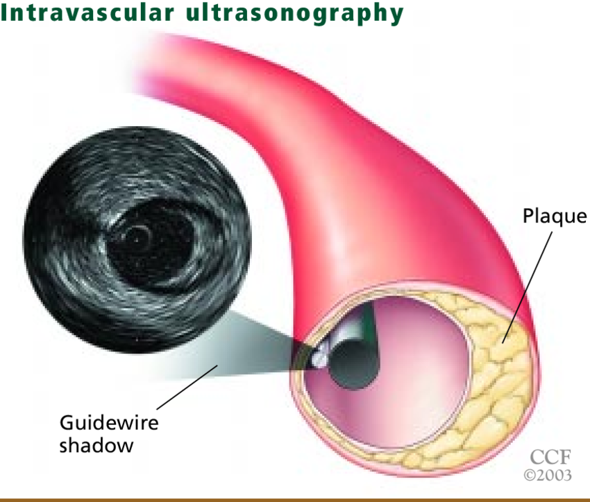
You will not feel the catheter  in your artery and you will not feel any pain during acquisition of the ultrasound images.

You may need to lie flat on your back with pressure applied to the catheter insertion site for a few hours after the test to or prevent bleeding. In some cases, your physician may use a device that seals the small hole in the artery, called a "closure device," which will allow you to move around sooner.
For several hours, your catheter site will be checked for bleeding or swelling and your blood pressure and heart rate will be monitored. Your physician may prescribe medication to relax your arteries to protect against spasm of the arteries or to prevent blood clots.

You may feel a little sleepy until the sedative has worn off

Your time in the hospital will vary depending on whether intravascular ultrasound was done in conjunction with another procedure such as catheter angiography or angioplasty. While intravascular ultrasound itself does not add to your recovery time,  catheter angiography recovery will require you  to stay in the hospital for observation for up to six hours. Angioplasty and vascular stenting recovery may require 12 to 24 hours.

After you return home, you should rest and drink plenty of fluids. Avoid lifting heavy objects and strenuous exercise at least 24 hours. It is strongly recomended that you quit smoking as this is a major cause of atherosklerosis.

The catheter insertion site may  be bruised and sore. If bleeding begin where the catheter was inserted, you should lie down, apply pressure to the site  and call your physician.

Call your physician immediately if you notice any change in the color of your leg, pain, swelling or warm feeling in the area where the catheter was inserted.

Intravascular ultrasound part 6


How is the procedure performed?

Intravascular ultrasound is done in the catheterization lab, also called the cath lab, usually in conjuction with angiography or angioplasty.

This procedure is often done on an outpatient basis. However, some patients may require admission following the procedure. Please consult with your physician as to wether or not you will be admitted.

You will be  positioned on the examining table.

You may be connected to monitors that track your heart rate, blood pressure and pulse during the procedure.

A nurse or technologist will insert an intravenous (IV) line into your hand or arm so that sedative medication can be given intravenously. Moderate sedation may be used. As an alternative, you may receive general anesthesia.

The area of your body where the catheter is to be inserted will be sterilized and covered with a surgical drape.

Your physician will numb the area with a local anesthetic.

A very small skin incisions made at the site.

A sheath is first inserted into an artery (usually in the groin) or vein. Using x-ray or ultrasound guidance, the catheter is inserted into the sheath and gently maneuvered through the vessel to the target location. Once in place, the transducer on the end of the catheter uses sound waves to produce pictures of the blood vessels. Doctor can move the catheter to get pictures of the inside of the vessels at the different locations.

At the end of the procedure, the catheter will be removed and pressure will be applied to stop any bleeding. The opening in the skin is then covered with a dressing. No sutures are needed.

Intravascular ultrasound part 5


How does the procedure work?

Intravascular ultrasound uses high-frequency sound waves to provide images from inside the blood vessels. Sound waves sent from the transducers bounce off of the walls of the artery and return to the transducers as echoes. A computer helps convert these echoes into images on the monitor to produce pictures of the blood vessels.

Intravascular ultrasound part 4


What does  the equipment look like?

The intravascular ultrasound catheter is thin, flexible tube with tiny ultrasonic transducer attached to one end. The other end of the catheter connects to a computer workstation that converts the sound waves from the transducer into real-time images on a monitor.

Intravascular ultrasound part 3


How should I prepare?

You should inform your physician of any medications being taken and if there are any allergies especially to iodinated contrast materials. Also inform your doctor about recent illness or other medical conditions.

You will be asked to remove some of your clothes and to wear a gown during the exam. You may be also be asked  to remove jewelry, removable dental appliances, eye glassess and any metal objects or clothing that might interfere with the x-ray images.

If sedation is used for the procedure, you may be asked not to eat or drink anything for four to eight hours before your exam. Be sure that you have clear instructions from your health care facility.
Do not drive for 24 hours after your exam if sedation is used. Arrange for someone to drive you home. Because an observation period is necessary following intravascular ultrasound and the exam is often used in conjunction with another procedure, you should ask your doctor if you will need to be admitted to the hospital overnight.

Intravascular ultrasound part 2


What are some common  uses of the procedure?

Intravascular ultrasound is most often used to visualize the coronary arteries in conjuction with or to help plan for catheter angiography or angioplasty and vascular stenting. Unlike angiography, intravascular ultrasound can show the entire artery wall and reveal more information about plaque build up (atherosclerosis). Information from intravascular ultrasound often affects treatment decisions, such as the sizing of a stent and where it should be placed. It is often used after angioplasty and vascular stenting to confirm the stent has been placed correctly and that the procedure has addressed the problem.

Intravascular ultrasound part 1


What is intravascular ultrasound?

Intravascular ultrasound uses a transducer or probe to generate sound waves and produce pictures of blood vessels.  Ultrasound does not use ionizing radiation, has no known harmful effects, and provides a clear picture of soft tissues that don't show up well on x-ray images. This procedure  requires little or no special preparation.
Intravascular ultrasound, also known as endovascular ultrasound or intravascular echocardiography, is an imaging technique that uses a transducer or probe to generate sound waves and produce pictures of the insides of blood vessels. The technique uses a special catheter with a small ultrasonic transducer on one end. The catheter is threaded through an artery or vein to the target location, where sound waves are generated by the transducer to produce images of the blood vessels and help physicians assess their condition.

Intravascular ultrasonography


Intravascular ultrasonography is relatively new technique capable of detecting subtle vascular abnormalities. Intravascular ultrasonography can give a 3 dimensional, 360º visualization of the vessel and can detect more vascular  abnormalities than can angiography.

Doppler techniques


Doppler techniques appear valuable in detecting stenosis in grafts. Percutaneous transluminal angioplasty performed under color Doppler ultrasonography guidance is useful in maintaining and improving graft patency. Percutaneous transluminal angioplasty with color Doppler ultrasonography guidance allows patients to avoid surgical intervention, hospitalization,  and adverse reaction to contrast media and exposure to ionizing radiation, with reduced cost  and with  better graft survival.

Arteriovenous (AV) graft infection part 9


CARE AFTER CRYOPRESERVED ALLOGRAFT PLACEMENT

Cannulation of cryopreserved allografts is possible 10-14 days after placement, and when swelling has subsided so that the course of the arteriovenous graft can be palpated. Aseptic technique during cannulation, including standard precautions for hand washing and glove changes, is recommended to minimize risk of access infection. Cannulation technique should be a hybrid of the techniques   for expanded polytetra fluoroethylene arteriovenous graft regarding depth of the access  and the texture of an autogenous vein. It is also necessary to rotate cannulation sites in order to avoid pseudoaneurysm formation. A retrospective study using constant cannulation (button hole technique) with cryopreserved femoral veins showed good outcomes related to patency and minimal infection risk.

A qualified individual should perform a physical examination to detect arteriovenous graft dysfunction at least monthly. The 3 preferred surveillance techniques for stenosis of arteriovenous graft are:
  • intra access flow using sequential measurements with time analysis
  • directly measured or derived static venous dialysis
  • duplex ultrasound

Other acceptable techniques include physical findings of persistent swelling of the arm, presence of collateral veins, prolonged bleeding after needle withdrawal, or altered characteristics of pulse or thrill in the arteriovenous graft. Unstandarized dynamic venous pressures should not be used.

Arteriovenous (AV) graft infection part 8


ALLOGRAFT METHOD VS GRAFT EXCISION METHOD

There are 2 methods for treating infected hemodialysis arteriovenus graft - the allograft method and the graft excision method. The graft excision method is generally used to manage a synthetic graft infection.
  • The allograft method is a single procedure which involves removing the infected arteriovenous graft and implanting the cryopreserved allograft in the same infected site. Preserving the vascular access saves potential future arteriovenous access site. Access is possible 10-14 days after implantation.

  • The graft excision method involves two separate procedures. First the infected arteriovenous graft is removed. After the infection has cleared, a new arteriovenous graft is placed in a different location, which diminishes potential sites for future access

A temporary central venous catheter and intravenous antibiotic therapy are needed for both methods. The duration of the central venous catheter  is generally longer for the graft excision method because a central venous catheter is needed after the first and  second procedures, until the new arteriovenous graft is ready for cannulation.

Arteriovenous (AV) graft infection part 7


CRYOPRESERVED ALLOGRAFT

Cryopreserved allografts are cryogenically preserved cadaver vessels.

Cryopreserved allograft is an option for treating infected hemodialysis arteriovenous graft. Allografts have been implanted either adjacent to or directly into the infected fields using the same anastomotic regions, thus saving other sites for future access.

Arteriovenous (AV) graft infection part 6


Total removal of infected arteriovenous graft and placement of  a new access device at a remote site may be required. This involves placement of a temporary dialysis catheter until the infection is resolved and results in losing the original graft site. Because potential access sites are limited, partial graft excision to salvage a site is also an acceptable method for treating graft infection.

Lastly, patient with forearm arteriovenous graft removed may have adequate upper arm veins for an arteriovenous fistula, or another expanded polytetrafluoroethylene graft may also be a consideration, since the time to cannulation can be as short as 24-hours post operatively.

Arteriovenous (AV) graft infection part 5


Arteriovenous graft infection management is a balance between resolving the infection while preserving the vascular access as follows:

SUPERFICIAL INFECTION
  • Treat initially with broad-spectrum antibiotic therapy to cover both gram-negative and gram-positive microorganisms
  • Base subsequent antibiotic therapy upon culture results
  • Incision and drainage may be beneficial

EXTENSIVE INFECTION
  • Treat with appropriate antibiotic therapy
  • Resection of the infected graft materia

Arteriovenous (AV) graft infection part 4


Infection prevention is critical for vascular access maintenance. Strict dialysis precaustions and aseptic technique are important in preventing and minimizing access infection. The World Health Organization recommends washing hands as follows:
  1. Before touching a patient
  2. Before clean/aseptic procedures
  3. After body fluid exposure risk
  4. After touching a patient
  5. After touching patient surroundings

Arteriovenous (AV) graft infection part 3


Gram positive organisms are the main cause of graft infections, with Staphyllococcus aureus being the most common arteriovenous graft-related bacteria. Infection caused by Staphyllococcus aureus prone to more complications and worse outcomes than other infections. Consequences of arteriovenous graft  infection include prolonged catheter-dependence leading to morbidity, multiple vascular-access procedures, loss of access site, lengthy hospitalizations, metastatic infection and patient mortality. Sepsis with multiple organ failure and endocarditis are other serous complications. One month prior to arteriovenous graft infrection, many patients may have a lower serum albumin level (albumin <3.5 mg/dl) - a well known mortality risk factor. Another consideration is occult infection of old non functioning arteriovenous graft, which has been linked to erythropoetin-resistant anemia and chronic inflammation.

Arteriovenous (AV) graft infection part 2


Risk of infection associated with arteriovenous graft includes insufficient antisepsis during surgical procedure, length of function, repetitive cannulation, venipuncture technique with risk of contamination or hematoma formation. Other risk factors are history of multiple infections, number of surgical revisions,obesity and thrombosed. Moreover, patients on dialysis have compromised immune function, and those with diabetes are more prone to infection and poor wound healing. Graft site also impacts infection risk. Access infection is more likely to occur in tigh grafts than in upper extremity grafts due to potential for the enteric organism contamination. For this reason, placement of arteriovenous graft in lower extremity site is ussually a last resort.

Arteriovenous (AV) graft infection part 1


Non-arteriovenous fistula access is a prime risk factor for infection. The synthetic material of arteriovenous graft, commonly expanded polytetrafluoroethylene, is porous and therefore provides an opportune medium for the formation of biofilms, causing general infection. Biofilms make the resident microbes resistant to both natural and pharmacologic defences. The incidence of arteriovenous graft infection is from 9% (forearm) to 20% (tigh). While this is significantly lower than central venous catheter, it is ten times higher than arteriovenous  fistula. Infection is a common cause graft loss.

The arteriovenous (AV) graft part 5


At each hemodialysis treatment:
  • Make sure that the nurses can see your access and needles at all times during your treatment
  • Unless instructed otherwise by your nephrology team, it is important that needles are put into your graft in different areas for each treatment
  • At the end of each treatment, the needles are removed. Apply light pressure for 10 minutes (or as instructed by your nephrology team) using the fingers from your other hand at each needle site to promote clotting at the site
  • If bleeding start again once you leave your dialysis unit, apply light pressure for 20 minutes and if bleeding does not stop, go to nearest emergency departement

The arteriovenous (AV) graft part 4


Routinely check for and report to your nephrology team  (surgeon, doctor or nurse) any of the following:
  • A change in colour, temperature or sensation (numbness or tingling) and pain in the hand  of your graft arm (or foot of your graft leg)
  • An increase in swelling, pain or redness on your arm (or graft leg) and the presence of any fluid leaking from your incision
  • Fevers, chill or loss of thrill
  • Bleeding that occurs from your graft when you are not in the dialysis unit
  • Needle sites that are not healing
  • Your surgeon or nephrology team member will give you other important instructions

The arteriovenous (AV) graft part 3


Surgery to create an arteriovenous (AV) graft

A surgical procedure is needed to create the arteriovenous graft. An anesthetist will talk to you about the anasthetic that will be best for your surgery.
After the surgery, its usually takes two to four weeks for the graft to be ready for use. Then, each time you have a hemodialysis treatment the dialysis nurse will insert two needles into your graft.

Care of your arteriovenous graft

After the surgery, follow your surgeon's instructions about your dressing and when the stitches (sutures) will be taken out.
Check for a "thrill" which is the sensation like a vibration caused by blood flowing through your graft and can be felt just above your incision line. The "thrill" indicates the arteriovenous graft is working.

The arteriovenous (AV) graft part 2


Benefits of an arteriovenous graft
  • A graft is usually the access method of choice if your veins are not suitable for a fistula
  • The graft can usually be used for hemodialysis two to four weeks after surgery
  • One the incision is healed, there are no restrictions regarding showering or swimming

Sunday, September 23, 2018

The arteriovenous (AV) graft part 1


The Arteriovenous (AV) graft

When the most common method access, the arteriovenous  (AV) fistula, cannot be made, the arteriovenous (AV) graft is a suitable substitute.
An arteriovenous graft is a strong artificial tube inserted by a surgeon underneath the skin of your forearm, upper arm or thigh. One end of the tube connects to one of your arteries, and the other end connects to one of your veins (in the same limb). The graft may be straight or in the shape of a horseshoe. The surgeon will decide whether to use your arm or leg for the arteriovenous graft. The decision depends on many factors including the size and condition of your veins and arteries. There is a higher risk for infection and clotting with an arteriovenous graft than with an arteriovenous fistula

Management of arteriovenous graft thrombosis part 3


Interventional thrombolysis

Prosthetic graft thrombosis can be treated with various percutaneous techniques and tools, including combinations of thromboaspiration, use of thrombolytic agents such as tissue plasminogen activator (tPA), mechanical thrombectomy  and mechanical thrombectomy devices. Independent of the applied technique it is important to perform thrombolysis as soon as possible to avoid the need for a central venous  catheter and as an outpatient procedure to decrease costs, whenever possible. Post procedural angiography to detect and correct inflow, intra-access or venous outflow stenosis is mandatory.

Management of arteriovenous graft thrombosis part 2


Surgical thrombectomy

Surgical thrombectomy is performed with a thrombectomy catheter. On-table angiography should be performed after completion of the arterial and venous limbs of the graft. This should visualize the central venous outflow as well as the graft, It is required to exclude residual thrombi and define the cause of thrombosis. Identification and simultaneous correction of the underlying stenosis are integral parts of any surgical or interventional declotting procedure.

Management of arteriovenous graft thrombosis part 1


Graft thrombosis should be treated without unnecessary delay and within 48 hours, at least before the next dialysis session. Early declotting allows for immediate use of the access without the need for a central venous catheter. There is always a compact "arterial plug" present. Old thrombi (>5days) are often fixed to the vessel wall beyond the venous anastomosis, making surgical extraction more difficult. This is less problem for the radiological treatment.

Management of arteriovenous graft stenosis part 2


Persistent stenosis

Some stenosis cannot be dilated by conventional balloon angioplasty. These "hard" stenosis can be treated with cutting ballons or ultra high pressure balloon (up to 32 atm)


Recurring stenosis

Recurring stenosis can be treated radiologically, with or without stent placement, or surgically. The strategy for treatment should be made considering the individual condition of the patient in relation to the invasiveness of the surgical treatment. In spite of complete opening of the percutaneous  transluminal angioplasty balloon of sufficient diameter, the dilated vessel wall may collapse immediately after removal of the balloon. This elastic recoil can be prevented  by stent implantation, especially in central veins. Stent placement in the needling areas of forearm fistula should be avoided except for percutaneous transluminal angioplasty-induced ruptures not controllable by protracted balloon inflation.

Management of arteriovenous graft stenosis part 1


A diameter reduction of >50% of the lumen together with a significant flow decline is considered as an indication for treatment.

Stenosis at the arterial anastomosis

As in autogenous fistula, most arterial inflow stenosis in grafts can succesfully be treated by percutaneous transluminal angioplasty. Stenosis of the arterial anastomosis itself can be dilated, if only the afferent artery and the graft at the anastomosis are affected and there is no stenosis in the efferent artery. If there is an additional stenosis of the efferent artery, angioplasty of the anastomosis alone will enhance graft flow with the risk of peripheral ischemia due to reduced peripheral arterial perfusion. In these patients, either dilatation of the efferent artery by interventional radiology through surgical revision of the anastomosis may resolve the dilemma.

Management of autogenous arteriovenous (AV) fistula thrombosis part 3


Surgical thrombectomy

Surgical thrombectomy is performed with thrombectomy catheter. Manual retrograde thrombus expression can be helpful. On-table venous outflow angiography of the recanalized vein as well as the central veins should be performed whenever possible to find/exclude additional stenosis  or residual thrombus. Identification and concurrent correction of the underlying causes of thrombosis are essential parts of any surgical or interventional declotting. The best results of surgery probabily will be encountered  after proximal re-anastomosis for anastomotic stenosis of forearm arteriovenous fistula, which is the most frequent location of stenosis  in this type of access. Primary patency of the new proximal anastomosis has been reported to be as high as 80% at 1 year and 67% at 2 years. If access failure recurs frequently in a short time period, a new fistula may need to be created.

Management of autogenous arteriovenous (AV) fistula thrombosis part 2


Interventional trombolysis

Trombolisis can be performed mecahanically or pharmacomechanically. While, the immediate
success rate is higher in grafts than autogenous arteriovenous (AV) fistula (99 vs 93% in forearm fistula), the primary patency rate of the forearm arteriovenous fistula at 1 year is much higher  . One year secondary patency rates are 80% in forearm and 50% in upper arm arteriovenous fistula., respectively. In arteriovenous fistula, the combination of a thrombolytic  agent (urokinase or tissue plasminogen activator=tPA) with balloon angioplasty resulted in  an immediate success rate of 94%. Flow restoration was achieved in 88,9% of the arteriovenous fistula.

Management of autogenous arteriovenous (AV) fistula thrombosis part 1


Fistula thrombosis should be treated as soon as possible or within 48 hours. The duration and site of arteriovenous fistula thrombosis as well as the type of access are important determinants of treatment outcome. Timely declotting allows immediate use without the need for a central venous catheter. Thrombi become progressively fixed to the vein wall, which makes surgical removal more difficult. Thrombosis may affect the post-anastomotic vein segment as result of anastomotic stenosis or may begin at the needle site. When the clot is localized at the anastomosis in radial-cephalic and brachial-cephalic fistula, the outflow vein may remain patent due to the natural side branches that continue to carry venous blood flow. In these accessess it is possible to create a new proximal anastomosis. Thrombosis in transposed basilic vein fistula usually leads to clot propagation of the entire vein. Although comparative studies are missing, the available literature suggests that thrombosed autogenous arteriovenous fistula should, preferably, be treated by interventional radiology. The single exception may be forearm arteriovenous fistula, thrombosed due to anastomotic stenosis. It is likely that in such cases, proximal re-anastomosis will provide good results.

Thursday, September 20, 2018

Management of autogenous arteriovenous (AV) fistula stenosis part 5


Persistent stenosis


Some stenosis cannot be dilated by conventional balloon angioplasty. These "hard" stenosis can be treated with cutting ballons or ultrahigh pressure ballons.

Recurring stenosis

Recurring stenosis can be treated radiologically, with or without stent placement, or surgically. The strategy for treatment should be made considering the individual condition of the patient in relation to the invasiveness of the surgical treatment. In spite of complete opening of the percutaneous transluminal angioplasty balloon of sufficient diameter, the dilated vessel wall may collapse immediately after removal of the balloon, This elastic recoil can be prevented by stent implantation, especially in central veins. Stent placement in the needling areas of forearm fistula should be avoided except for percutaneous transluminal angioplasty-induced ruptures not controllable by protracted balloon inflation.

Management of autogenous arteriovenous (AV) fistula stenosis part 4


Balloon angioplasty

In order to visualize the stenosis, angiography is performed by retrograde puncture of the brachial artery, in case of anastomotic problems, or by direct antegrade puncture of the vein above the anstomosis if an outflow problem is suspected. It is controversial, wether long-segment stenosis should be treated radiologically or surgically. While some authors recommend surgical intervention, either by graft interposition or vein transposition, others recommend radiological intervention. Studies proving the superiority of one of two treatment options for the treatment of long-segment obtructions are not available. However, percutaneous transluminal angioplasty of short-segment stenosis  (<2cm) has a better outcome compared with long -segment stenosis (>2cm)

Management of autogenous arteriovenous (AV) fistula stenosis part 3


Venous outflow stenosis

Percutaneous transluminal angioplasty (PTA) is the first treatment option in the outflow veins (cephalic/basilic). Junctional stenosis,  of the superficial veins  with the deep venous system, can also be treated by percutaneous transluminal angioplasty. If a stent is placed in the final arch of the cephalic vein, it must not protude into the subclavian vein where it could induce stenosis and preclude future use of the distal (basilic, brachial and axillary) veins.

Management of autogenous arteriovenous (AV) fistula stenosis part 2


Stenosis of the anastomotic area

Surgical treatment is indicated in stenosis of the anastomotic area located in the lower forearm. Alternatively, percutaneous transluminal angioplasty (PTA) is possible although its results are likely to be less long-lasting. Primary interventional treatment is indicated in stenosis of the anastomotic area located in the upper forearm and in the upper arm. Surgery should be considered in cases of early or repeated reccurences of the lessions. Dilatation or surgical revision of anastomotic stenosis in upper arm fistula can cause steal syndrome and access-induced hand ischemia. Careful dilatation up to 5 or 6 mm initially is recommended. Dilatation to >6 mm is rarely indicated